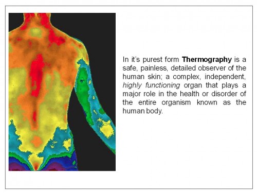
Thermography is a 100% safe imaging test that can reveal a road map to improve your health, wellness and life longevity. Thermography is non-invasive and painless, involves no radiation, no contact, is private and F.D.A. Certified.
Thermography is a screening device for the purpose of assisting the health practitioner to care for their patients by promoting health and prevention of disease by using the physiological warning signs made possible by thermography, or infrared imaging. Thermography may detect changes that if left unattended, could possibly progress into a late stage disease that could then be detected by other types of imaging devices.
Unlike X-Ray, sonography, magnetic resonance imaging (MRI) and virtually all other modern medical imaging modalities designed to capture anatomical information, medical Infrared technology is designed to capture physiological information.
Digital Infrared Thermal Imaging, better known as Thermography, is an FDA certified adjunct to (not replacement for) mammography. Thermography is not an x-ray and there is no touching or compression of the body. Thermography is a test of physiology that uses a very sensitive medical digital infrared-sensing camera that develops a color image on a computer of a body’s thermal patterns. The underlying principle by which Thermography could show certain kinds of disease is because tumors have an increased blood flow and increased number of blood vessels to maintain their increased cellular growth. This causes an increase in temperature that is seen on the thermographic scan. Thermography is the only modality that can demonstrate whether a condition called estrogen dominance is affecting the breasts. Estrogen dominance is a potential risk factor for the development of breast cancer, fibrocystic breasts, uterine fibroids and ovarian cysts. Mammography does not indicate hormonal imbalance. Thermography can.
However, skin temperature also is affected by larger blood vessels and the metabolic character of tissues deeper in the body. These energy features are transmitted to the skin surface and can greatly influence its temperature. The energy features are characteristically diffused by depth and non-homogeneity of the underlying tissue. While characteristics of skin perfusion are of interest in Thermology studies of the peripheral nervous system, it is the vascular and metabolic features of deeper tissue that is of principle interest to a study.
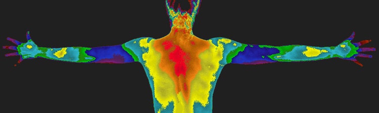
Modern imaging devices provide a way for physicians to instantly capture large arrays (images) of quantitative thermographic information. Each pixel represents a different temperature. Imaging is available for full body or a specific area (s) of interest. A prescription is not necessary.
Facts:
- Safe, easy , pain-free, and radiation-free
- Provides a color coded temperature “fingerprint”
- No contact, compression, or pain
- Detects physiological changes of the body in real time imaging
- Identifies inflammatory conditions by temperature changes
- Identifies fibrocystic tissue disease and tumor inflammation within the body
- Effectively and safely screens dense breasts and women with implants
- Useful for evaluating chest wall size after breast surgery
- Effective for breasts of all sizes
- Creates opportunities for early intervention!